A comprehensive eye exam is a detailed evaluation of your vision and the health of your eyes. It involves a series of tests to assess different aspects of your visual system. During an exam, an eye care professional looks at various structures within the eye to detect any potential issues. Here’s information on eye exams and what eye structures are observed during them:
1. The Conjunctiva
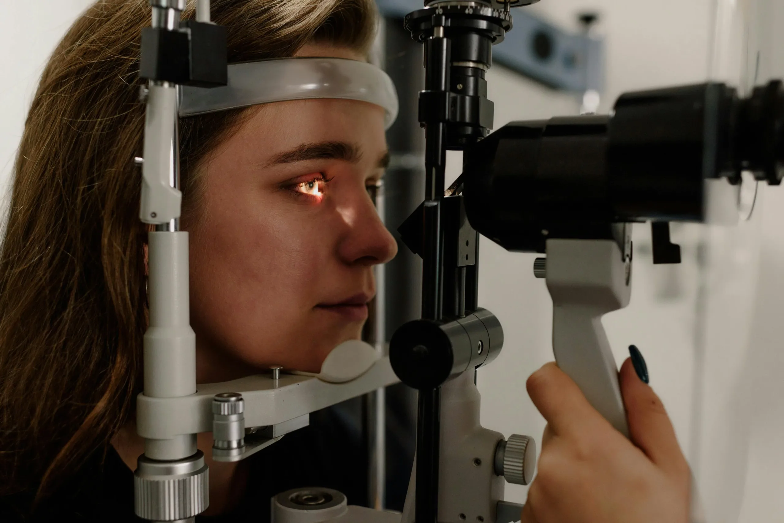
The conjunctiva is the thin, clear membrane that covers the white part of your eye, known as the sclera, and lines the inside of your eyelids. It plays a role in lubricating the eye and protecting it from foreign particles. To examine the conjunctiva, an optometrist typically uses a slit lamp, which is a microscope with a bright light. This instrument allows for a highly magnified view of the eye’s surface structures.
What Does the Doctor Look For?
An optometrist will look for signs of inflammation, like redness or swelling. Eye inflammation could indicate conditions like conjunctivitis (pink eye), allergies, or dry eye. The blood vessel’s health within the conjunctiva is also assessed, as changes can sometimes point to systemic health issues.
How Do Routine and Medical Eye Exams Differ?
While both routine and medical eye exams are necessary for maintaining ocular health, they address different needs. A routine eye exam is primarily preventative, focusing on the overall health of the eyes and the correction of vision issues, like:
- Nearsightedness
- Farsightedness
- Astigmatism
Medical eye exams are diagnostic in nature, aimed at identifying, monitoring, or treating specific eye diseases or systemic conditions that affect vision. Medical exams are generally prompted by symptoms like sudden vision changes, eye pain, or conditions like diabetes and hypertension that impact eye health.
2. The Iris
The iris is the colored part of your eye that surrounds the pupil, which is the black opening in the center. The iris controls the amount of light that enters the eye by adjusting the size of the pupil. Your doctor will examine the iris and pupil using a penlight or the slit lamp. They then check the pupil’s response to light, observing whether it constricts properly and equally in both eyes. The shape, size, and color of the iris are also observed for any irregularities that might suggest underlying conditions.
3. The Cornea
The cornea is the transparent, dome-shaped outer layer at the very front of the eye. It covers the iris, pupil, and anterior chamber, and it is responsible for most of the eye’s focusing power. The anterior chamber of the eye is the fluid-filled space between the cornea and the iris. Eye doctors closely observe the anterior chamber during an examination to assess its depth, clarity, and the presence of any abnormalities.
A detailed examination of the cornea is conducted with the slit lamp. Your optometrist carefully inspects the surface for any scratches, abrasions, or opacities. Assessing the cornea’s clarity and curvature is key, as conditions like corneal abrasions, infections, or diseases like keratoconus can affect vision quality.
4. The Optic Nerve
The optic nerve is a bundle of more than a million nerve fibers located at the back of the eye, responsible for transmitting visual information. It transmits data from the retina to the brain, allowing you to see. To view the optic nerve, the doctor may use an ophthalmoscope or the high-magnification lenses of a slit lamp.
Your pupils may be dilated with special eye drops to provide a wider, clearer view of the back of the eye. The doctor assesses the optic nerve’s color, size, and shape. Examining this nerve is beneficial for detecting signs of glaucoma, a condition characterized by damage to the optic nerve, which can lead to vision loss if not managed.
5. The Retina
A retina is the light-sensitive tissue that lines the back of the eye. It captures light and converts it into electrical signals that are sent to the brain through the optic nerve. Using a slit lamp, usually after pupil dilation, the optometrist examines the retina for any signs of disease or damage. They will check the health of the retinal blood vessels and look for abnormalities that could indicate conditions like:
- Diabetic Retinopathy
- Macular Degeneration
- Retinal Detachment
Schedule an Eye Exam
Regular eye examinations provide a detailed look at the complex structures of your eyes. Having these key parts of the eye evaluated allows for the early detection and management of potential issues. Contact an optometrist today to schedule your next comprehensive eye exam.