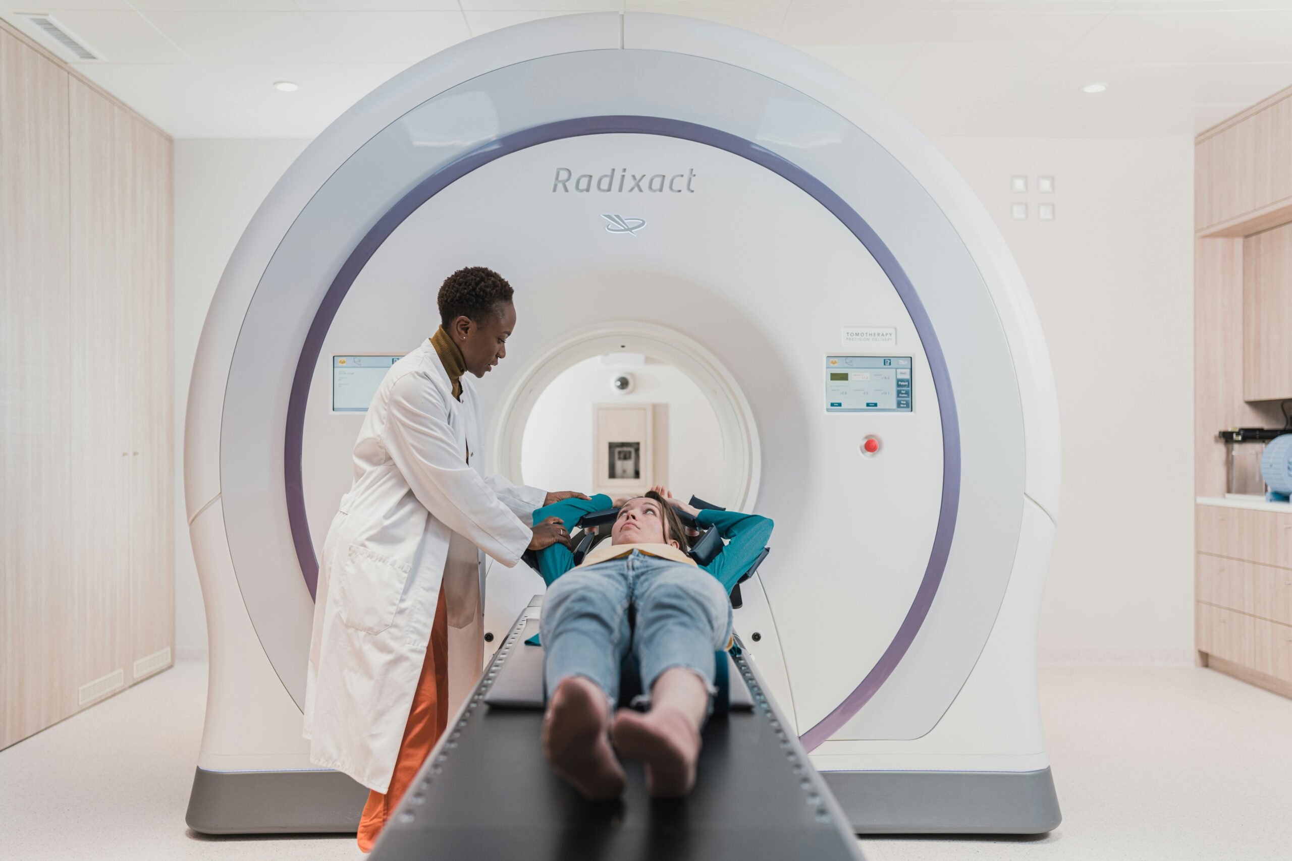
Magnetic resonance imaging (MRI) is a visualization method used in radiology, and it uses radio waves to generate pictures of the body’s organs and tissues. While you may have heard of it, understanding how it functions can help you be more prepared. Here is more information about the role of MRI technology in modern medicine:
Creating Detailed Pictures
MRI machines produce detailed images of soft tissues. These pictures help physicians examine parts of the body, and these areas may not be as visible with other imaging techniques. An MRI scan does not use ionizing radiation, which is found in X-rays and CT scans.
These detailed scans provide a clear view of organs and structures. For some people, an MRI of the abdomen shows the liver, gallbladder, and pancreas with great clarity. Physicians use these images to assess the condition of these internal organs, and it helps guide treatment.
Evaluating Blood Vessels
A specific type of MRI, called magnetic resonance angiography (MRA), focuses on blood vessels, and this creates images of arteries and veins to check blood flow. If your doctor needs to look closely at your vascular system, they may suggest an MRA. It is a noninvasive way to see blood vessels without needing surgery.
Identifying Vein Problems
Another specialized MRI is used to look at veins, and this procedure specifically creates images of the venous system in the body. If a physician needs information about venous circulation, they might use an MRV, which is tailored to highlight veins.
While an MRI is used to evaluate veins in the head and neck, it can also be applied to other areas of the body, such as the legs or torso. The detailed images from this technique help doctors assess the structure and function of the veins. This information supports them in understanding your specific situation. Typical areas of focus include:
- Veins within the skull
- Veins in the abdomen
- Veins in the legs and pelvis
Diagnosing Spinal Issues
Magnetic resonance imaging is a standard tool for examining the spine. When you have back or neck pain, an MRI may be used to get a clear picture of what is happening. The scan shows the spinal cord, nerve roots, and surrounding tissues in detail, and it provides a different view than an X-ray, which mainly shows bones.
An MRI of the spine can show several kinds of tissue. It can visualize the discs between your vertebrae, and it also shows the ligaments that hold them together. Doctors review these images to identify irregularities, which might be related to your symptoms.
This detailed imaging is helpful for many spinal conditions. Because the scan provides such a clear view of soft tissues, it is useful for looking at disc-related issues. The information from the scan gives your doctor a clearer understanding of your spine’s condition, and it guides treatment courses.
Schedule Magnetic Resonance Imaging Today
MRI technology provides detailed views of the body’s internal structures. From soft tissues and blood vessels to the spinal cord, it gives doctors a comprehensive look inside the body. If your doctor has recommended an MRI, specialists can help you schedule it. Contact a diagnostic imaging center today to book your appointment.