Hydrocephalus is a condition characterized by an abnormal accumulation of fluid in the brain, affecting patients of all ages. Accurate diagnosis and tailored treatment plans are key to managing this condition effectively. Neuroradiology plays a central role in identifying and evaluating hydrocephalus, using advanced imaging technologies to provide precise insights. This article examines how neuroradiology aids in diagnosing hydrocephalus, highlights effective imaging techniques, and explores how scans inform treatment plans.
How Does Neuroradiology Diagnose Hydrocephalus?
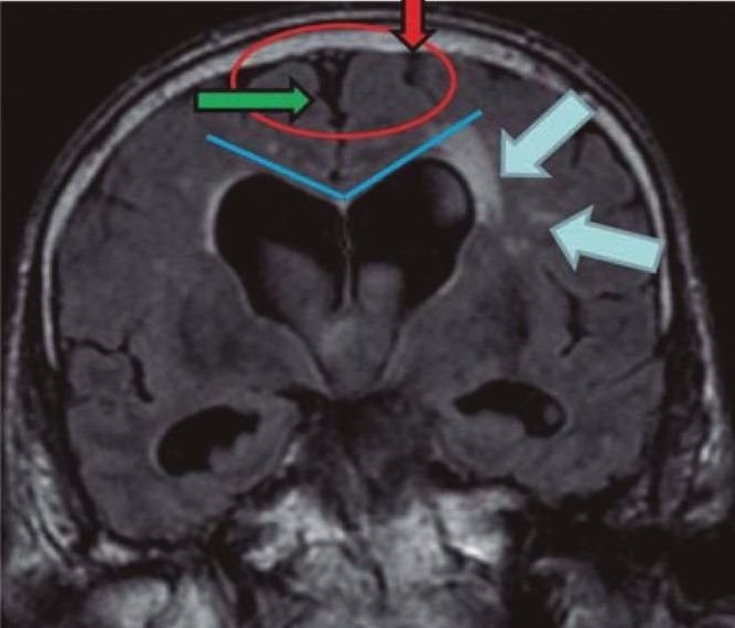
Neuroradiology uses a range of imaging techniques to assess hydrocephalus. These methods enable healthcare professionals to measure fluid levels within the brain and identify structural changes that may indicate potential complications. The process often begins with assessing physical symptoms to determine whether imaging is necessary.
Once the need for imaging is established, neuroradiologists utilize tools such as magnetic resonance imaging (MRI) and computed tomography (CT) scans to produce detailed images of the brain. These scans enable them to detect enlargement of the ventricles or other irregularities that indicate fluid buildup. By analyzing these images, specialists gather the necessary data to recommend treatments or further tests.
Which Imaging Techniques Are Most Effective?
Various imaging modalities are used in neuroradiology, each offering unique advantages. The following are commonly utilized techniques with their applications:
- Magnetic Resonance Imaging (MRI): MRI provides detailed images of soft tissue, making it ideal for assessing fluid levels and structural abnormalities in the brain. It is particularly useful for identifying the source of fluid buildup.
- Computed Tomography (CT) Scans: CT scans offer rapid imaging and are often used in emergency settings to detect acute cases of hydrocephalus. Though less detailed than MRI, they are effective for identifying enlarged ventricles.
- Ultrasound (for infants): Through a fontanelle ultrasound, specialists can assess brain structures in infants without exposing them to radiation. This method is commonly used when hydrocephalus is suspected in younger children.
- Nuclear Medicine Scans: By using a small amount of radioactive material, nuclear medicine scans can assess cerebrospinal fluid dynamics. These scans help trace fluid pathways to detect obstructions.
Each technique plays a role in forming a complete understanding of a patient’s condition. Based on the findings, specialists decide which method serves the diagnostic or treatment purpose best.
How Do Scans Guide Treatment Plans?
Neuroradiology not only aids in diagnosis but also helps design treatment strategies tailored to the specific needs of the patient. The insights gathered inform surgical and non-surgical approaches for managing hydrocephalus.
Surgical Planning
Imaging guides surgeons in identifying the best course of action for managing hydrocephalus. For cases requiring shunt placement, neuroradiology pinpoints the optimal site for insertion by visualizing fluid buildup and pathways. Advanced scans might also assist in endoscopic third ventriculostomy (ETV), a minimally invasive procedure sometimes used as an alternative to shunt systems. By using these imaging results, surgeons can improve the precision of interventions.
Monitoring and Follow-Up
After treatment, neuroradiology continues to play a part in monitoring the outcome. Follow-up scans verify that shunts or other interventions are functioning properly, while also tracking the patient’s recovery. Periodic imaging can detect any recurrence of fluid buildup, enabling adjustments to treatment as needed. This ongoing role of imaging underlines its contribution throughout the treatment process.
Consult a Specialist
If hydrocephalus is suspected, consulting a specialist is necessary for accurate diagnosis and care. Neuroradiologists use advanced imaging technologies to assess the condition and develop a personalized treatment plan. Timely intervention can significantly improve outcomes and reduce potential complications. With advancements in neuroradiology, patients now have access to better diagnostic tools and treatment options. Reach out to a specialist to address concerns and explore the best path forward.