Ultrasound is a medical imaging technique that uses high-frequency sound waves to create images of the inside of the body. These sound waves, produced by a handheld device, travel through the skin and bounce off internal organs and tissues. Since the technology does not use radiation, it offers a distinct method for observing various anatomical structures. Here is more information about the role of ultrasound imaging:
Providing Dynamic Visualization
Different ultrasound techniques provide various types of information. One common approach produces two-dimensional images that help evaluate the size and texture of internal structures. These images are useful for monitoring changes in certain medical conditions over time. Advanced techniques enable the visualization of blood flow in real-time, allowing clinicians to assess your health.
Another technique focuses on assessing blood flow by analyzing how sound waves interact with moving cells. This method helps visualize the direction and speed of blood movement, aiding in the evaluation of vascular health and identifying potential issues like blockages or abnormalities. By combining these advanced imaging techniques with other diagnostic tools, clinicians can develop comprehensive treatment plans.
There are methods designed to measure tissue stiffness. These methods, which can change due to disease, work differently. By sending vibrations through the tissue and analyzing their movement, this approach provides a non-invasive way to assess structural changes, offering valuable insights into the progression of certain conditions.
Diagnosing Conditions
Providers may use ultrasound to diagnose a range of conditions and to monitor their progression. Other applications include monitoring thyroid nodules and evaluating joint inflammation in rheumatoid arthritis. There are various types of chronic conditions that are monitored regularly.
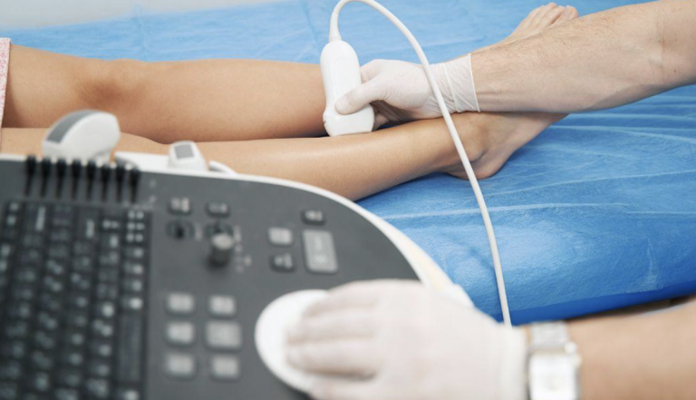
Showing Tissues as They Move
Ultrasound uniquely captures real-time images of tissues and organs in motion. This dynamic quality is especially valuable when assessing musculoskeletal conditions. Providers can watch muscles contract and tendons glide, so they are able to identify abnormalities that might not be visible in a static image.
This real-time feedback is also beneficial during certain guided procedures. A physician performing a biopsy can watch the needle’s path on the ultrasound screen, which allows for precise targeting of a specific area while avoiding adjacent structures. This visual guidance enhances the accuracy of tissue sample collection, leading to better pathological examination results.
Including Clinic or Home Use
The portability of modern ultrasound systems allows for their use in various settings beyond a hospital’s radiology department. Compact, high-quality ultrasound machines are now common in outpatient clinics and private medical offices. Because the equipment is more accessible, it facilitates more frequent monitoring for patients with chronic illnesses without requiring a hospital visit. Some portable systems are even being explored for home use under professional supervision, offering a potential path for remote patient management.
Schedule an Ultrasound Today
Ultrasound provides a non-invasive method for observing internal body structures, visualizing blood flow, and assessing tissue movement to support the ongoing management of chronic health conditions. Its use in various clinical settings makes it a versatile tool for both initial diagnosis and long-term monitoring. If your healthcare provider recommends this imaging, contact a diagnostic center and schedule your ultrasound appointment.