
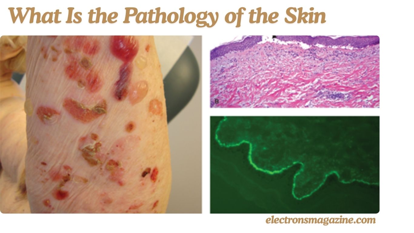
The skin is a complex organ that protects the body, controls temperature, prevents water loss, and helps detect touch and pain. When skin tissue becomes abnormal because of disease, infection, inflammation, or cancer, skin pathology helps doctors understand what is happening.
In simple terms, the pathology of the skin is the study of abnormal changes in skin cells and tissues. This article explains how skin pathology works, what doctors look for, how biopsies are examined, and why pathology is important for diagnosing different skin conditions.
What Is the Pathology of the Skin?
The pathology of the skin is the study of diseases and abnormal changes that affect skin tissue. It looks at what happens to skin cells, layers, blood vessels, hair follicles, glands, and other structures when a disease develops.
Doctors can sometimes identify a skin condition simply by examining its appearance. However, some conditions look very similar on the surface. In these situations, a skin biopsy may provide additional information.
A small piece of affected skin is removed and sent to a laboratory. A pathologist or dermatopathologist examines the tissue under a microscope and looks for changes that may help identify the condition.
Skin pathology can help identify:
- Inflammatory skin diseases
- Skin infections
- Autoimmune conditions
- Benign skin growths
- Precancerous changes
- Skin cancers
- Some hair and nail disorders
- Certain rare skin conditions
The main purpose is to understand how the tissue has changed and what those changes may mean for the patient.
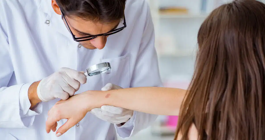
Why Is Skin Pathology Important?
Skin pathology is important because not every skin condition can be accurately diagnosed by appearance alone.
A red patch, lump, sore, or changing mole can have many possible causes. A dermatologist may have a strong idea about the diagnosis, but a biopsy can provide additional evidence when the diagnosis is uncertain.
Pathology can help doctors:
- Confirm a suspected diagnosis
- Rule out other conditions
- Identify abnormal cell growth
- Diagnose certain skin cancers
- Understand unusual inflammation
- Identify some infections
- Determine whether a growth is benign or malignant
- Guide further medical treatment
It can also prevent unnecessary treatment. If two skin conditions look similar but require different treatments, identifying the correct condition is important.
The value of skin pathology comes from connecting microscopic findings with the patient’s symptoms, medical history, and physical examination.
What Are the Main Layers of the Skin?
Understanding normal skin structure makes it easier to understand skin pathology.
Alos Read: What Is Dermatology Practical and Conceptual? Everything You Need to Know
The skin is commonly divided into three major layers:
- Epidermis
- Dermis
- Subcutaneous tissue
Each layer has its own role and contains different types of cells and structures.
Epidermis
The epidermis is the outermost layer of the skin. It provides much of the body’s protective barrier.
It contains mainly keratinocytes, which develop and mature as they move toward the surface of the skin.
The epidermis also contains specialized cells involved in pigmentation, immune defense, and sensation.
Abnormalities in the epidermis can cause changes such as:
- Scaling
- Thickening
- Peeling
- Abnormal pigmentation
- Changes in cell growth
- Certain types of tumors
Dermis
The dermis is located below the epidermis.
It contains collagen, elastic fibers, blood vessels, nerves, hair follicles, sweat glands, sebaceous glands, and other structures.
Changes in the dermis can cause swelling, inflammation, scarring, changes in blood vessels, and abnormalities around hair follicles and glands.
Subcutaneous Tissue
The subcutaneous layer lies beneath the dermis.
It contains fat and connective tissue and helps cushion the body, store energy, and provide insulation.
Some skin diseases and tumors can extend into this deeper layer, which is why the depth of a biopsy can be important.
What Does a Dermatopathologist Examine?
A dermatopathologist is a specialist who studies diseases of the skin and related tissues under a microscope.
When examining a skin biopsy, the specialist may look at the size, shape, arrangement, and behavior of cells.
They may also examine:
- The thickness of the epidermis
- The condition of the dermis
- Inflammatory cells
- Blood vessels
- Hair follicles
- Sweat glands
- Sebaceous glands
- Pigment
- Abnormal growth
- Tissue damage
- Tumor cells
The overall pattern is very important.
For example, simply finding inflammation does not automatically identify a particular disease. The specialist needs to determine what type of inflammation is present, where it is located, and what other changes occur around it.
Clinical information also helps the dermatopathologist interpret the biopsy correctly.
How Does a Skin Biopsy Help Diagnose Skin Disease?
A skin biopsy provides a sample of tissue that can be examined more closely.
Doctors choose the biopsy method according to the suspected condition and the part of the skin that needs to be examined.
Common types include:
- Shave biopsy
- Punch biopsy
- Incisional biopsy
- Excisional biopsy
A shave biopsy removes a relatively superficial portion of skin.
A punch biopsy removes a small circular section of skin and can provide tissue from several layers.
An incisional biopsy removes part of a larger lesion.
An excisional biopsy removes the complete lesion when appropriate.
The goal is to collect a representative sample.
The doctor may consider the size, location, appearance, and suspected cause of the skin problem when choosing the most suitable method.
What Happens During Skin Pathology Testing?
After a biopsy is taken, the tissue is sent to a pathology laboratory.
The sample must be carefully preserved and prepared before it can be examined.
In a routine process, the tissue is preserved, processed, embedded, and cut into very thin sections. These sections are placed on microscope slides and stained so that different structures can be seen more clearly.
The pathologist then examines the slides.
A common stain used for routine tissue examination is hematoxylin and eosin, often called H&E.
Depending on the case, additional tests may be needed. These can include special stains, immunohistochemistry, immunofluorescence, or other laboratory methods.
The testing process depends on what the doctor and pathologist are trying to determine.
What Is Skin Histopathology?
Skin histopathology is the microscopic examination of skin tissue to identify changes caused by disease.
Healthy skin has an organized structure. Disease can change that organization.
A histopathology examination may show:
- Increased skin thickness
- Abnormal cell growth
- Inflammation
- Changes in pigmentation
- Blister formation
- Tissue destruction
- Abnormal blood vessels
- Changes around hair follicles
- Changes in connective tissue
- Tumor development
Histopathology is particularly useful when the clinical appearance does not provide enough information to make a confident diagnosis.
However, microscopic findings must be interpreted carefully because several diseases can sometimes produce similar patterns.
What Are the Common Pathology Patterns of Skin Diseases?
Dermatopathologists often identify specific reaction patterns in skin tissue.
These patterns are not always the final diagnosis. Instead, they help organize the possible causes.
Common patterns include spongiotic, psoriasiform, interface, lichenoid, granulomatous, and vesiculobullous patterns.
Spongiotic Pattern
A spongiotic pattern involves increased fluid between cells in the epidermis.
It can occur in several types of dermatitis and eczema.
The microscopic pattern may help support a diagnosis, but additional clinical information may be needed to determine the exact cause.
Psoriasiform Pattern
A psoriasiform pattern involves changes in epidermal growth and thickness.
Psoriasis is one condition that can produce this type of pattern.
The immune system plays an important role in psoriasis and can cause increased inflammation and faster production of skin cells.
Interface Pattern
An interface pattern involves changes around the area where the epidermis meets the dermis.
It can occur in several inflammatory and immune-related skin diseases.
The exact diagnosis depends on the details of the microscopic findings and the patient’s clinical presentation.
Vesiculobullous Pattern
This pattern is associated with vesicles or blisters.
Microscopic examination can help show where separation has occurred within the skin.
Some blistering disorders require additional testing to determine whether immune proteins are present.
How Does Inflammation Appear in Skin Pathology?
Inflammation is one of the most common findings in skin pathology.
Alos Read:How Can I Find Dermatologos Cerca de Mi Ubicación? Complete Guide
The immune system produces inflammation when the body responds to infection, injury, irritation, or other triggers.
A pathologist may examine which inflammatory cells are present and where they are located.
Common inflammatory cells include:
- Lymphocytes
- Neutrophils
- Eosinophils
- Plasma cells
- Histiocytes
- Mast cells
The type and location of these cells can provide useful diagnostic clues.
However, inflammation does not automatically mean that a particular disease is present.
Different conditions can produce similar inflammatory responses. Therefore, the overall microscopic pattern and clinical history are important.
How Are Skin Infections Diagnosed Through Pathology?
Skin infections can be caused by bacteria, fungi, viruses, or parasites.
Some infections can be recognized from characteristic changes in the tissue. In other cases, additional laboratory tests may be necessary.
A pathologist may look for:
- Microorganisms
- Changes in skin cells
- Tissue damage
- Characteristic inflammatory patterns
- Viral changes
- Fungal structures
- Other evidence of infection
Special stains can sometimes make certain organisms easier to identify.
The best testing method depends on the suspected infection.
Pathology may also be combined with cultures, molecular testing, or other laboratory methods when necessary.
How Does Skin Pathology Diagnose Skin Cancer?
Skin pathology plays an important role in diagnosing skin cancer.
Three major types of skin cancer are:
- Basal cell carcinoma
- Squamous cell carcinoma
- Melanoma
A suspicious lesion may be biopsied so that the tissue can be examined under a microscope.
Basal Cell Carcinoma
Basal cell carcinoma is a common type of skin cancer.
It develops from cells with basal characteristics and often grows slowly.
Pathology can identify the characteristic arrangement and appearance of the tumor cells.
Squamous Cell Carcinoma
Squamous cell carcinoma develops from keratinocytes.
Microscopic examination can identify abnormal squamous cells and help determine whether the growth is cancerous.
The pathology report may also provide information that helps doctors plan treatment.
Melanoma
Melanoma develops from melanocytes, which are pigment-producing cells.
Some melanocytic growths are harmless, while others are malignant.
Because melanoma can potentially spread to other parts of the body, accurate pathological examination is very important.
The pathologist may evaluate the structure and characteristics of the lesion and use additional tests when needed.
What Are Immunohistochemistry and Immunofluorescence in Skin Pathology?
Immunohistochemistry and immunofluorescence are specialized laboratory techniques used in selected cases.
Immunohistochemistry uses antibodies to detect specific proteins in tissue.
It can help answer questions about the type or origin of cells.
It may be useful when a tumor or other lesion is difficult to classify using routine microscopy.
Immunofluorescence is particularly useful for certain immune-related skin diseases.
It can detect immune proteins deposited in the skin and may help support the diagnosis of selected autoimmune blistering conditions.
These tests are not automatically required for every biopsy. They are selected according to the clinical question being investigated.
Which Skin Conditions May Require a Biopsy?
Not every skin condition requires a biopsy.
Many common conditions can be diagnosed through a physical examination and medical history.
A biopsy may be considered when:
- The diagnosis is uncertain
- A lesion looks unusual
- Several conditions look similar
- Skin cancer needs to be ruled out
- An inflammatory disease is difficult to identify
- A growth has changed
- A lesion does not respond as expected to treatment
- A rare condition is suspected
Conditions that may sometimes require pathology include unusual forms of eczema, psoriasis, autoimmune diseases, infections, benign tumors, precancerous lesions, and skin cancers.
The decision depends on the individual patient and the appearance of the lesion.
What Can Affect the Accuracy of a Skin Pathology Diagnosis?
Several factors can affect the quality and usefulness of a pathology result.
Biopsy Location
The sample should come from an area that represents the condition accurately.
Biopsy Depth
Some diseases affect deeper parts of the skin. A superficial sample may not contain enough tissue to identify the problem.
Biopsy Technique
The correct technique depends on the suspected diagnosis.
Timing
Skin lesions can change over time, so the stage of the lesion can affect its microscopic appearance.
Previous Treatment
Medications and topical treatments can sometimes change the appearance of diseased skin.
Clinical Information
Providing accurate information about symptoms, location, duration, medications, and suspected diagnoses can help the pathologist interpret the tissue.
These factors show why skin pathology works best as a partnership between the clinician and the pathology specialist.
How Is a Skin Pathology Report Interpreted?
A skin pathology report may contain several sections, including the specimen description, microscopic findings, diagnosis, and comments.
The final diagnosis is usually the most important part.
Some reports may use terms such as benign, malignant, inflammatory, atypical, dysplastic, or suspicious.
These words should be interpreted in their full medical context.
For example, an abnormal finding does not automatically mean cancer.
If you receive a pathology report that contains unfamiliar medical terms, discuss the complete report with the healthcare professional who ordered the biopsy.
Useful questions include:
- What is the exact diagnosis?
- Is the lesion benign or malignant?
- Does the lesion require additional treatment?
- Was the entire lesion removed?
- Do I need another biopsy?
- What follow-up is recommended?
Understanding the complete report is more useful than focusing on one unfamiliar word.
Alos Read: How Much Do Mohs Surgeons Make? U.S. Salary and Pay Guide
Frequently Asked Questions About Skin Pathology
What does pathology mean in dermatology?
Pathology in dermatology means studying abnormal changes in the skin caused by disease, infection, inflammation, immune problems, injury, or abnormal cell growth. It may involve examining a skin biopsy under a microscope.
Is dermatopathology only used for skin cancer?
No. Dermatopathology is used for many conditions, including inflammatory skin diseases, infections, autoimmune disorders, benign growths, unusual rashes, hair disorders, and skin cancers.
Does an abnormal skin biopsy always mean cancer?
No. An abnormal biopsy can show inflammation, infection, a benign growth, precancerous changes, or cancer. The exact diagnosis in the pathology report is more important than the word “abnormal.”
How long does it take to get a skin pathology result?
The timing varies depending on the type of biopsy and whether additional testing is needed. Simple cases may be completed sooner, while complicated cases requiring special tests or expert review can take longer.
Can skin pathology diagnose a skin condition without a biopsy?
Some skin conditions can be diagnosed clinically without a biopsy. However, when the diagnosis is uncertain or a suspicious lesion needs evaluation, a biopsy may provide important additional information.
Conclusion
The pathology of the skin is the study of abnormal changes that occur in skin cells and tissues because of disease, inflammation, infection, immune reactions, injury, or abnormal growth.
It is an important part of dermatology because many skin conditions can appear similar on the surface. A biopsy and microscopic examination can reveal details that cannot always be identified through a visual examination alone.
Skin pathology can help diagnose inflammatory diseases, infections, autoimmune conditions, benign growths, precancerous changes, and skin cancers. The most accurate diagnosis usually comes from combining microscopic findings with the patient’s symptoms, medical history, and physical examination.
If you have a skin biopsy or pathology report, it is best to discuss the complete result with your dermatologist or qualified healthcare professional. A single medical term should not be used to make a diagnosis without considering the full clinical picture.